Wednesday, December 10, 2014

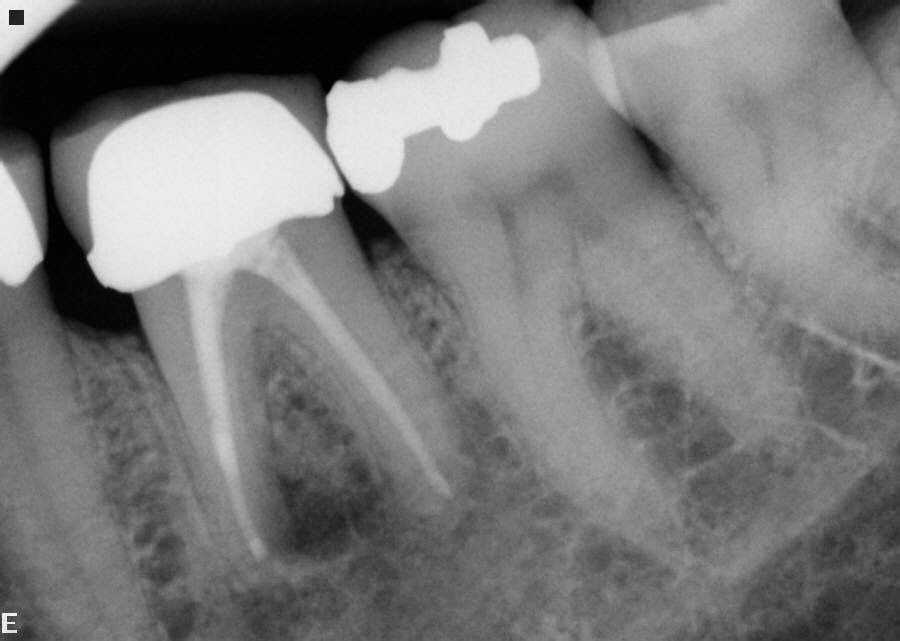
Case 41 Lateral canal with apical curve

Pre operative x-ray #4

Patient was symptomatic.


Post operative x-ray #4


Note: Two lateral canals and curvature negotiated and obturated.


Comments: When contemplating root canal therapy on any tooth, curvatures can be problematic.  It is an ideal place for instruments to separate when the canal system is really small, this is a case that should be referred.  It was fortunate in this case the lateral canals were filled and a radial lucency can clearly be seen on a mesial aspect of this root. 

Wednesday, December 3, 2014

Case 40 Curvature at the Apex

Pre-operative x-ray #31 with
Positive/Negative reversed
Pre-operative x-ray #31
Patient was symptomatic and options were discussed; apicoectomy vs retreatment. Retreatment was the preferred option due to the location of the tooth.


Post - operative x-ray #31
Post-operative x-ray #31 with
Positive/Negative reversed


 Please note curvature of the apex.


Comment: The patient was symptomatic probably due to incomplete cleaning and obturation of the last 4-5mm of the mesial root which was extremely curved. With judicious cleaning of the mesial root and negotiation to the apex, a good fill is now noted. Prognosis is excellent.

Thursday, November 13, 2014

Case 39 Lateral canals found in the funniest places

Pre-operative x-ray #6


Large apical lesion on the mesial aspect of #6. Buccal tissue is swollen and patient is asymptomatic.


Immediate post-operative x-ray #6


Obturation completed. Note lateral canal on the mesial aspect of the tooth. 


6 mo. post-operative x-ray #6


Patient is asymptomatic and lesion is practically gone. Please note bifurcation at apex also. 


10 mo. post-operative x-ray #6


Lateral lesion has practically healed completely.


Comments:  Lateral canals are often the reason that periapical lesions are present or teeth have continual problems after root canal therapy.  In many cases, they are not seen until after the work is completed. If a lesion remains after a root canal has been done and does not heal, many times unfilled lateral canals are the cause.

Thursday, November 6, 2014

Case 38 2 canals often in distal root

Pre-operative x-ray #30


Patient was symptomatic and swollen.


File film #30


Second distal canal was located. 


Immediate post-operative x-ray #30


Retreatment was completed. Mesial canals were also retreated.


10 month check-up x-ray #30


Complete healing after ten months. 


Comments:  Always a choice between a surgical approach and retreatment on any root canal that is failing, if one is assured that a fracture is not the problem. Generally retreatment will confirm that either the canals have been poorly cleaned or other canals that have never been negotiated are present, as in this case of the distal root.

Wednesday, October 22, 2014

Case 37 Five canals more common than we think

Pre-operative x-ray #19

Patient presented with swelling and painful to bite.


Photo showing pulp chamber floor #19


Once the pulp chamber was clear, five canals clearly visible. Note: The fifth canal is usually in the isthmus area between distal buccal and distal lingual systems, if in the distal root. 


Post-operative x-ray #19


All canals were filled to the apex.


Comments: Usually if there are five canals you will find three in the mesial root more often than the distal root. Three canals in the either root are usually not three separate canals.  In my experience, a third canal either joins the  buccal or lingual canal system.  I can tell you from surgically treating first molars, that the isthmus area many times is the cause of periapical problems because the isthmus has not been or cannot be cleaned adequately.

Tuesday, October 14, 2014

Case 36 Lateral canals can be important.

Preoperative x-ray #4


Tooth is symptomatic.


Immediate post operative x-ray #4


Obturation with 2 lateral canals.


2 month post operative check #4


Tooth is asymptomatic.


6 year post operative x-ray #4


Note: resorption of lateral canal filling material.  Probably just sealer only, gutta percha does not dissolve. Lateral lesions have healed.
Please note: Apical lesion on #5.


7 year post operative xray


All lesions have healed around #4 and no evidence of lateral canals being filled.  Root canal was completed on #5.


Comments:
If lateral canals are not filled i.e. cleaned, there is a good chance that a tooth's apical area will never heal. 

In most cases, it is impossible to mechanically cleanse an lateral canal system. However, sodium hyperchloride may chemically clean a lateral canal. In most of my cases, if a lateral canal is filled it is with softened gutta percha and sealer.  This is speculation on my part, however. 

Wednesday, October 1, 2014

Case 35 Apicoectomy vs. Orthograde Retreatment


Pre-operative x-ray #19

Root canal completed by another provider with separated file in ML canal.  Tooth is extremely symptomatic.


Post-operative x-ray #19


Surgical approach was decided upon and no fractures noted. Three retro fills were placed, and a separated file was removed from ML canal with ultrasonics.  Despite large size of lesion and the amount of bone loss, the prognosis was good. 


3 month check-up #19

Patient is asymptomatic and healing was noted.


9 month check-up #19

Patient continues to be asymptomatic and the bone almost completely filled in


Comments:  Separated instruments are just a way of life for those who do root canal therapy.  The instrument itself is not a problem.  What it represents is: a blockage of a possible dirty canal.  In this case, I just speculated that the separated instrument was the cause of this patient's problem.  Do I know this for sure? No.  Therefore all canals were retrofilled.