Wednesday, May 28, 2014

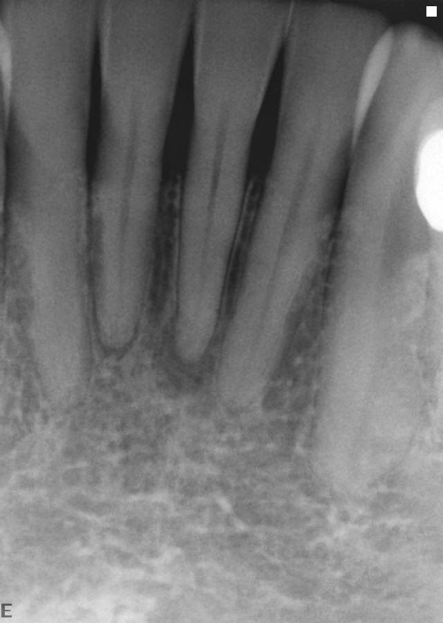
Case 28 Think outside the box: buccal approach

Pre-operative x-ray #24
Top view of access #24


Lingually displaced lower anterior
Straight line approach with file #24
Post-operative x-ray #24


Final filling in place #24

Comments:
Lower anteriors, especially when displaced to the lingual, are more readily accessed on the buccal instead of the lingual. This is by far the easiest way to approach root canal therapy when aesthetics is not an issue. The bonded composites on the market provide matching color to the surrounding enamel.

Wednesday, May 21, 2014

Case 27 Age 101 and counting


Pre-operative x-ray #12,13

I performed root canal on #12 when the
patient was a young man in his early 90's.

Patient presented with symptomatic tooth # 13
 with no radio-graphic visible canal spaces.


Post-operative x-ray #13


Comments:
It would seem to me that I am treating more and more older (65+) patients who present not only with health problems, but with very small sclerosed canal systems.  My best advice is if you are contemplating doing a root canal on a senior citizen or anyone where canal spaces are not clearly visible on an x-ray, I would suggest not attempting at all. Experience has taught me that at one time each tooth has a canal(s) and it takes less time each time I work on these kind of teeth because I have a little better idea of where the canal orifices should be.


Wednesday, May 14, 2014

Case 26 Separated instrument - surgical approach

Pre-operative x-ray #13


Separated lentulo-type paste filler in apical third of root.
Periapical lesion present and the tooth is symptomatic to occlusal pressure.


Photo of removed instrument with bur for scale


Post-operative x-ray #13


Comments:
Separated instruments pose two problems: should they try to be removed or should the tooth holding the separated file be surgically retreated? My philosophy is any separated file in the top one-third of the canal system has a good chance of being removed. I also believe any separated instrument in the last half of the canal system is usually non-retrievable. If a surgical approach is done, all attempts should be made to retrieve the separated instruments apically. Any kind of apical filling seals best when a metal instrument has been removed. It is difficult to make an adequate apical preparation against a metal instrument. Indecently, plastic thermal files are relatively easy to prep against.

Wednesday, May 7, 2014

Case 25 Lateral canals - many times the key to success

Pre-operative x-ray tooth #5 


Patient presented with symptomatic #5.  X-ray shows lateral lesions on both roots, one to the distal and one to the mesial.


Post-operative x-ray #5

Note: both lateral radiolucensies


Lateral canals in both roots


14 month post-operative x-ray #5

Tooth is asymptomatic and lateral radiolucensies are healed.


Comment: Lateral lesions on teeth with no evidence of fracture usually indicate a lateral canal system. If root canals are done and the lesions fail to heal, I would generally retreat before contemplating other options, i.e. surgery or extraction.
Wednesday, April 30, 2014

Case 24 Surgical Approach Multiple Teeth

Pre-operative x-ray

Periapical abscess approximately 2cm across with de-vital #22 & #25.  Root canals on 23 & 24 were completed some time in the past. Patient is symptomatic.


Immediate post-operative x-ray


Non negotiable canals in both #22 & #25.  No attempt was made to try to negotiate the canal space in #22 as there was no obvious canal visible. #22-25 were retrofilled.

4 month post-operative x-ray


Bone fill in is very apparent and all symptoms are gone.


Comments: 
Lower anteriors are very difficult to treat and their premature loss poses a very difficult restorative situation. In this instance, it was decided that retreatment of #23 & #24 would not be beneficial in the long term and a surgical approach would be more predictable as far as healing was concerned.

Wednesday, April 23, 2014

Case 23 When a fracture is not a fracture, tooth #18

Pre-operative x-ray


Necrotic tooth with periapical lesions


Photo of access opening with fracture circled in red


Please note: fracture line down distal
Immediate post-operative x-ray


6 month post-operative x-ray


Periapical radiolucencies are gone


Comment:
When fractures are noted, careful probing of the pockets in the area of the fracture line is needed.  Many times a good periapical x-rays will show bone loss which will indicate a non-restorable tooth. Patients need to be informed of fractures and the possibility of eventual tooth loss if one does exist. However, in my experience, teeth that are periodontally sound after initital root canal treatment and immediate full coverage tend to remain so.

Wednesday, April 16, 2014

Case 22 Why root canals fail/ 5 canals, tooth #19


Pre-operative x-ray


Previous root canal treatment.  It appears that there are spaces in the root canal system that have not been negotiated or cleaned, especially the distal root.


Post-operative x-ray

Note: 3 files in distal root


Photo: 5 canal orifices
Post-operative x-ray


All canals were cleaned and filled to the radiographic apex.


Comments:
Though teeth may have multiple canal systems and some of the canals may meet apically, a dirty canal will inevitably cause periapical symptoms as contamination may occur from the unclean canal.  A canal obturated with gutta percha will not stop contamination from a neighboring canal that joins it.