Wednesday, March 26, 2014

Options: retreatment versus a surgical approach


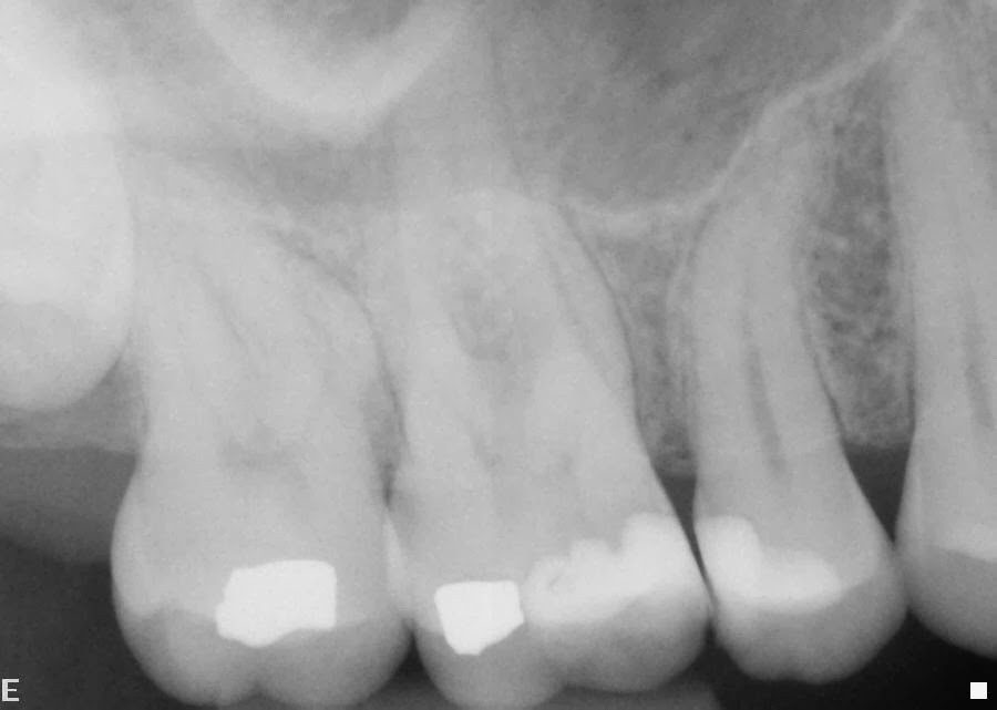
Post operative x-ray 9 months after root canals on #27,28 


Original root canal was completed in my office.  Patient was asymptomatic but radiolucent areas were still present.  Patient desired no additional treatment.


1 year post operative x-ray


Same as 9 months


Pre-operative x-ray / 6 year check up


There was a significant change in radial lucency and the tooth was becoming symptomatic.


Immediate post-operative x-ray


Surgical approach was decided for both #27 and #28 as being the most predictable as the original root canal therapy seemed to be adequate.  During the surgery no fractures were found nor any unusual periodontal problems.


15 month check up


Patient is asymptomatic and apical healing is observed around both roots.


Comments:
It is always difficult to decide whether to retreat a root canal or to approach it surgically. If all canals have been found and the obturation seems to be adequate, probably a surgical approach would be more appropriate and more predictable.


Thursday, March 20, 2014

Case 18: Predictable endodontics

Case 18a: pre-operative x-ray
Case 18a: photo


Case 18b: pre-operative x-ray

Case 18b: photo


Please note in both cases alloy on pulp chamber wall.


Comments:
As noted above many restorations are placed close to if not into the pulp chamber. In both of these cases, patients were very surprised that this was the case. Granted, most of the time the dentist who place the restoration tell patients of the situation and the possible need of root canal therapy due to the depth of the filling.  However, in my experience, patients never hear this. If a restoration will probably lead to future root canal therapy, it needs to be well documented in the patient's record and explained in detail to the patient as to not infer it was the dentist's "fault" it was so deep.  Most of the time, these fillings are replacements for other fillings because of fracture or decay. Symptoms may occur only after the new filling is placed.  It also needs to be explained that the tooth may be asymptomatic prior to the new filling, but may become symptomatic after the filling.

Wednesday, March 12, 2014

Case 17 Fractured tooth

Pre-operative x-ray #3


Tooth was symptomatic, sensitive to chewing and biting.


Photo #3 after filling material removed


Note: fracture line extending from the mesial to the distal, making the tooth non restorable.


Comment:
Any tooth suspected of a non restorable fracture should be investigated with an access opening and removal of all restorative materials so that the pulp chamber and walls of tooth can easily be exposed. This will make it much easier to diagnose whether or not the tooth is restorable.

Wednesday, March 5, 2014

Case 16 Anterior tooth with internal resorption repaired with MTA

Pre-operative x-ray showing internal resorption
No history of trauma according to the patient. The tooth was asymptomatic.  Patient was not aware of the problem until it was picked up on by the general dentist doing routine x-rays.


1 week post-operative x-ray


No perforation was noted. Periodontally, probings were within normal limits.


3 year post-operative x-ray


Tooth is sound and patient is asymptomatic.


Comment:
MTA is the material of choice for internal resorption.  If you can probe into the resorbed area (in other words a perforation has occurred) MTA will not work.  Also note that many times it is extremely difficult to negotiate a canal system to the radiographic apex.  In this case the system that I was able to clean was entirely filled with MTA.  I would suggest a series of  routine 6 month post op exams when teeth are treated in this manner. If internal breakdown continues to occur, probably extraction is the only alternative.  Patients need to be aware on their first appointment.

Wednesday, February 26, 2014

Case 15 Surgically treating primary teeth

Pre-operative x-ray primary tooth K
immediate post-operative x-ray

Patient presented with a symptomatic periapical lesion.


7 month post-operative x-ray

Comments:
Patient had a relatively new crown and we discussed possible retreatment.  However, it seemed that a surgical approach would probably be the most appropriate with the best results.  Patient understood if a fracture was present, the tooth would be removed.  It is obvious from the x-ray that crown-root ratio is very important and has to be taken into consideration when surgically treating these kind of teeth. Every attempt was made to keep the crown-root ratio the same while removing the apical lesion and placing retrofills.

Wednesday, February 19, 2014

Case 14 - Reimplantation as option to orthograde/surgical treatment tooth #18

Post-operative x-ray #18

Pre-operative x-ray #18

       
Five year follow up x-ray #18

Comment: Patient was very symptomatic and the root canal had been done some years previous. The anatomy was such that a surgical approach was not optimal due to a very broad buccal plate. The patient had TMJ problems which made an orthograde treatment difficult at best with a poor prognosis. My first thought was the possibility of a fracture, though there were no periodontal problems.  The time involved in removing a tooth, placing retrofills and re-implantation is about 5 minutes.  At the five year follow up the patient was asymptomatic and the tooth was firm with no periodontal problems.  The radiolucency at the apex was completely healed. The PDL space seems to have regenerated.

Thursday, February 13, 2014

Case 13 Exposing the pulp chamber

Pre-operative x-ray #19


When patient first presented for treatment. Patient was very symptomatic and told me dentist could not find all the canals.


Photo before treatment


Temporary that was in place removed, pulp chamber not touched

Please note: "material" fills mesial half of pulp chamber????


Photo after treatment


Pulp chamber fully exposed all canals easily identified.


Comment:
All canals need to be readily seen in order to be cleaned properly. The floor of the access opening must be large enough to demonstrate all three or four canals. Please note: The pulp floor is a different color than the walls of the access opening. Follow the color and you will find the canals.