Wednesday, August 17, 2016

Case 70 Three Definitive Mesial Canals

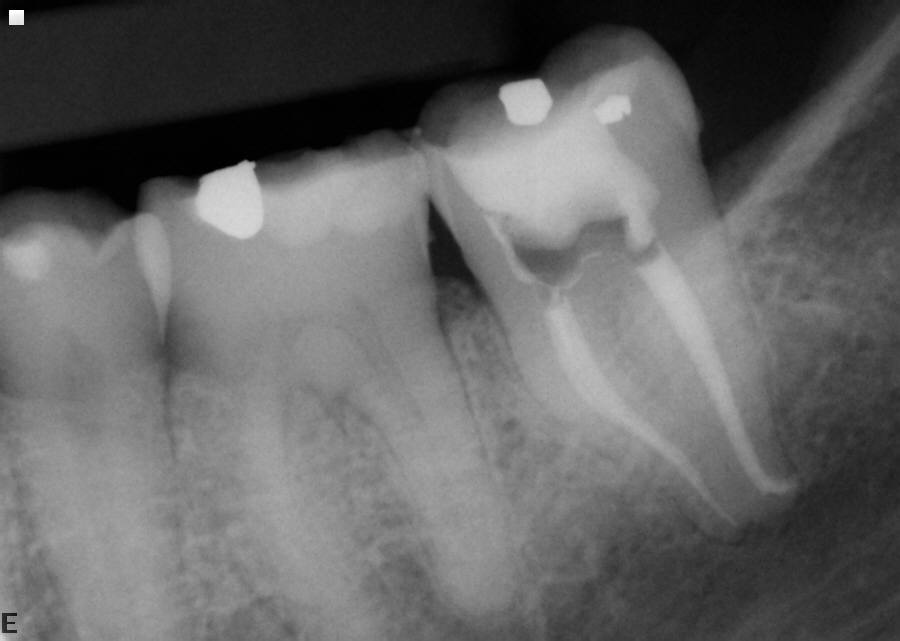
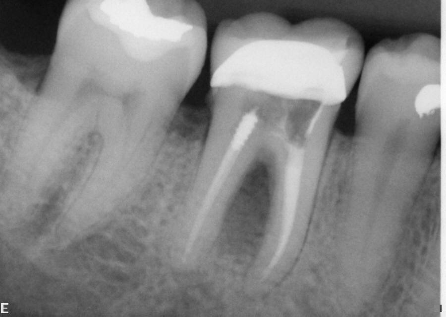
Pre-Op Xray #30


Post-Op Xray #30 (polarized xray)


Comment: Need I say anything?
Usually a "3rd canal" goes into either the mesial lingual or mesial buccal canals on a lower molar.
Occasionally, there are 3 separate canal systems in the mesial root of lower molars.
If you have properly cleaned the mesial lingual and mesial buccal canals and they are dry,
but there is exudate between the two, it usually means a 3rd canal is present.


Case 69 First Impression Fracture - Wrong!

Pre-Op Xray #4


Post-Op Xray #4


Post-Op Xray #4 (30 months)


Comment: If the periodontal situation is within normal limits, re-treatment may solve what appears to be a tooth that needs to be extracted, fractured??


Tuesday, August 16, 2016

Case 68 Bridge Salvaged

 
Pre-Op Xray #31


Post-Op Xray #31


Post-Op Xray #31 (6 months)


Comment: Extreme bone loss does not necessarily mean tooth loss. Proper root canal therapy can salvage a difficult situation.


Case 67 This Is Why We Re-treat


Pre-Op Xray #19


Post-Op Xray #19


Post-Op Xray #19 (13 months)


Comment: If there is any question about restoring a tooth that has less than ideal results, I think a consultation is required. Re-treating a tooth can effectively retain the tooth with complete apical healing.


Case 66 Good Result

Pre-Op Xray #31


                                                                               Post-Op Xray #31


Post-Op Xray #31 (8 months)


Comment: What you want to see, post operatively, in a necrotic tooth with periapical lesions.


Case 65 Tough Anatomy

Pre-Op Xray #17


Post-Op Xray #17


Comment: 3rd molars are difficult to treat. Not only because of the problem of actually getting the tooth isolated, but the root structure tends to be unusual ie: curved. This case is a good demonstration of the curvature of tooth #17.


Case 64 Re-treatment

Pre-Op Xray #18


               Post-Op Xray #18

 
Post-Op Xray #18 (11 months)


Comment: If a tooth has had a root canal which looks acceptable, yet symptoms remain, one of the options the patient must have is re-treatment. Then if the symptoms dissipate, one can assume that healing has started to occur as this case demonstrates.


Case 63 Excellent Result

Pre-Op Xray #31


Post-Op Xray #31

  
Post-Op Xray #31 (7 months)

 
Comment: If a lesion tends to be on the lateral side of a root, there tends to be a lateral canal besides an apical canal.


Case 62 What I Strive For

Pre-Op Xray #18


Post-Op Xray #18


Comment: Even though roots are curved ideal obtruation can be done.


Case 61 Metal Filling

Pre-Op Xray #11


Post-Op Xray #11


Comment:  Metal file used as filling material decades ago, now symptomatic. The file was removed (blackened) and the canal space cleaned to the apex. Gutta percha filling completed and temporary filling placed.  All symptoms disappeared.


Tuesday, December 8, 2015

Case 57 Mesial Buccal Root Amputation

Pre Op # 14

Fractured mesial buccal root. Patient had option to extract with implant or mesial buccal root amputation.


Final Film #14

Mesial buccal root removed. Solid bone around distal buccal and palatal roots.


Post Op 7 Months #14

Healing has occurred with bone fill in in the area of the mesial buccal root. Probing is within normal limits.


Post Op 13 Months #14

13 Months with crown. Pt extremely happy. No periodontal problems.


Comment: Some patients just do not want teeth removed. If within reason there are alternative treatments for fractured teeth, for instance, if a fractured root can be removed with no undue periodontal problems and the patient understands the home care situation, then amputation is a viable option. The patient must understand that a new crown has to be placed.

Case 60 MVA

Pre Op #22,23,24

22,23,24 are symptomatic to pressure, no response to cold.


Final Film #22,23,24

Asymptomatic


Post Op 8 Months #22,23,24

Asymptomatic, almost complete healing.


Comment: Oral trauma can devitalize teeth and one must test all teeth in a quadrant or a specific area to make sure a tooth that is necrotic is not overlooked.

Case 59 What Would You Do?

Xray #8,9

Horizontal fracture apical third.


Comment: Patient traumatized tooth #8 as a child years ago. Has been referred for possible root canal although there are no symptoms. Tooth responds slightly to an ice pencil but not near as does #9. The tooth is yellower in color than 9. My recommendation is no treatment, what is yours?

Case 58 The Perfect Root Canal?

Pre Op #30

Symptomatic #30.


Final Film #30

Ideal.


Comment:I wish they would all turn out like that.

Case 56 Locate ALL Canals


Pre Op #30

Symptomatic tooth, #30 that had been referred where all the canals had not been found. In particular, the mesial buccal canal had never been negotiated.


Pre Op #30

Additional Pre Op film


Final Film #30

Mesial buccal found and there were two canals in the distal.


Final Film #30

Additional final film.


Comment: When dealing with lower molars remember there are usually at least three canals. Two canals maybe found occasionally in second and third molars.

Monday, December 7, 2015

Case 55 Bone Loss in Furcation Usually Indicates Fracture, USUALLY

Pre Op #30
Root canal with furcation bone loss but not probeable.


Final Film #30

Retreatment completed, no fractures noted in access opening.


Post Op 6 Years #30
Bone completely fills furcation.


Comment: A dirty canal system in a tooth with a root canal is not recognizable on an x-ray. Cleaning the canal system is the only way to ensure that the canals are clean and sterlie. An access opening also can reveal the presence of a fracture many times.

Case 54 Retreatment Very Common, Mesial Root Not Cleaned

Pre Op #19
Symptomatic with swelling over mesial buccal root.


Final Film #19

At First appointment mesial canals cleaned thoroughly, at second appointment all canals were obturated down to radio graphic apex.


Post Op  21 Months #19

Symptoms gone and apex area of mesial root completely filled in with bone.


Comment: Mesial canals of lower molars are very commonly not cleaned well enough to the radio graphic apex. When retreatment done the canals in the mesial root are cleaned thoroughly down to the radio graphic apex.

Case 53 Third Molars Are Tough

Pre Op #32
Symptomatic #32 with Periapical lesion.


Final Film #32

Final Film


Post Op 13 Months #32
Periapical lesion gone.


Comment: Third molars can be tricky besides being hard to get to with a rubber dam. Don't be surprised if you tackle a RCT on a third molar that there may be a variety of canal systems from 1-5.

Case 52 Sclerotic Canals in Mesial Root

Pre Op #31
Symptomatic to pressure, necrotic.


Final Film #31
Final Film


Post Op 14 Months #31
Complete healing at 14 months.


Comment: Examine x-ray before starting. If canals can not be seen you probably don't want to start.