Thursday, December 19, 2013

Case #7 Orthograde Vs. Apicoectomy

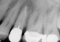
Pre op #10


Dentist attempted to locate the canal, could not.  Advised patient a possible  apicoectomy may be necessary.


File film #10 polarized view


Using ultrasonics I located the canal and  cleaned.  A polarized view will sometimes more clearly define the end of the file to the apex of  the tooth.


Immediate post op #10 polarized view


Again, this polarized view clearly  demonstrates the relationship of the root  canal filling to the apex of the tooth.  Please note: lateral incisors typically have a curve in  the apical end of the root.


Comments: Even though canals cannot be  initially negotiated, they are always there to  some degree. It is just a matter of finding  them.  Granted, some canals are not  negotiable, and surgery may be necessary. Generally surgery is not the first choice in root  canal therapy.
Wednesday, December 11, 2013

Case 6 Apicoectomy of 2 Teeth

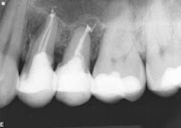
Pre-op #12 &13 previous root canal therapy


Patient was very symptomatic and buccal tissue slightly swollen, both teeth were very tender to touch and pressure.

Options were discussed ranging from exodontia to retreatment to apical surgery.  I recommended that the most predictable outcome would be apical surgery on both teeth.


Immediate post-op #12 & 13 after apicoectomy w/retrofills


No fractures were noted and periodontally there were no issues.


3 month follow up xray


Apical healing on both teeth was observed and the patient was completely asymptomatic.


Comment:
Questions always arise as to why things are not healing. In this case, is it the over-extension of the filling material? I think probably not. Usually it comes down to the fact the tooth canal system has not been properly cleaned or due to the anatomy of the canal systems cannot be cleaned well enough. Once the apical foramen is sealed, the symptoms disappear and healing can occur.

Thursday, December 5, 2013

Case #5 Two Separated Files w/Apical Lesion


Pre Op - 2 separated files in #13


Patient presented with 2 separated files and an apical lesion.  Patient was symptomatic. Options were discussed ranging from retreatment to a surgical approach to extraction. Patient did not want to loose the tooth and I recommended a surgical approach which would have a more predictable prognosis.


Immediate Post Op - Partial separated files with retrofill


Portion of the separated file in the facial canal system is present as well as the file in the lingual canal.  Retrofills were placed that covered both separated files and the isthmus between. 


8 month Check-Up


Patient is now asymptomatic and the apical lesion has completely healed.


Comment: Most separated instruments are impossible to remove especially if the breakage occurs in the apical third of the canal system. If the apical foramen can be successfully sealed the chances of success are greatly enhanced. It is necessary, however, to  have these patients on a routine 3-6 month recall for 2-3 years.

Wednesday, November 20, 2013

Case #4 separated file

Pre op xray separated in file in place


File was separated during cleaning of the canal.  Evidently it could not be removed or practitioner did not have the instruments to remove it.


Final post op completed RCT
Please note slight curve at the end of the root with a lateral canal going toward the distal.
Magnified view of post op apex


Comment:
Separated instruments in the upper third of the root system can generally be removed with ultrasonics. Separated files further down the system, especially around curves, are just about impossible to remove.  It is not the file that causes post operative problems,but rather, a canal system that was not cleaned properly and blocked by the file.

Wednesday, November 13, 2013

Case #3 retreatment #19 on 11 year old patient

Pre op: tooth #19 with a failing root canal
Please note the (underwhelming) root canal filling.  Patient, 11 years old, was quite symptomatic.

A young tooth is difficult to clean and obturate well because of the size of the canal system. 


Post op: completed retreatment


An alloy was placed in the access to ensure against any bacterial products.


3 year post operative x-ray


Periapical lesion has healed and tooth is  asymptomatic.

I recommend full coverage at 18 years of age to allow for structural changes in her dentition. Prior to 18 it is probably best that a posterior tooth have some sort of temporary crown.  I always recommend this to the patient and parents.


Tuesday, November 5, 2013

Case #2 Tooth #30 showing multiple silver points


Recommendation: Before a new crown is fabricated for a tooth that has had a root canal, making sure that the root canal is asymptomatic, no periapical lesions, and the root canal itself is of good quality. Additionally, all silver cone root canals should be retreated before new crowns are made.

This is an example of what I found in a lower first molar.
Pre Op - with Silver Points


Photo of ten silver points removed from tooth #30


Please note how corroded each point is.


Post Op - Root canal completed with gutta percha


(I can only speculate as to what the distal overhang is on the crown.)


Thursday, October 10, 2013

Case #1 Pre-op x-ray showing separated file
 CASE #1
This case was referred for retreatment or apicoectomy of Tooth #31.  On probing after anesthesia, it appeared that #31 was probably fractured.  There was a 9mm plus pocket on the mesial aspect of the mesial root.

Digital x-ray showed separated file at mesial root.
Case #1 x-ray of extracted tooth


Tooth was removed, dye placed over mesial root making the fracture visible.

Case #1 FX visible with dye
Case #1 Apex view w/File


Also note the separated file at the end of the mesial root.